Overview
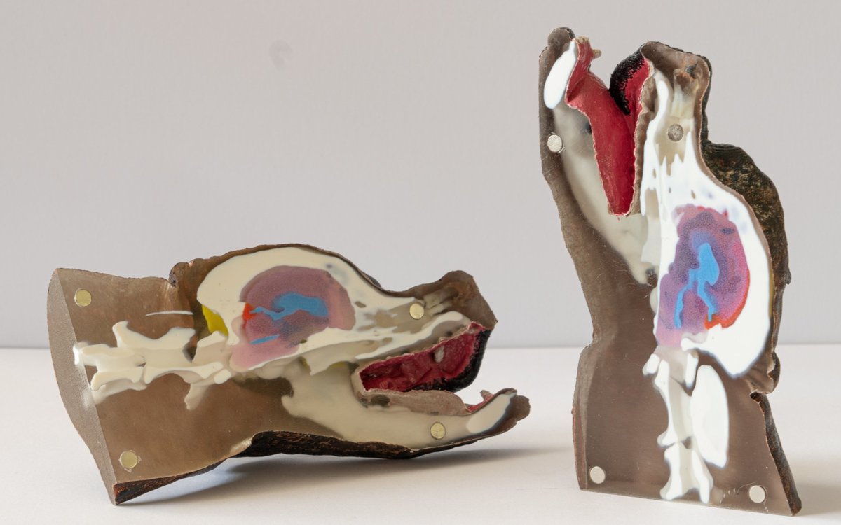
A collaboration with PennVet which aims to improve tools for canine neurosurgery, and to develop better real-time navigation and guidance for surgeons using patient-specific models.
Objective
Intracranial procedures such as tumor biopsy and resection remain technically demanding in veterinary neurosurgery, and effective patient-specific tools for planning and training are still lacking. Cadavers are commonly used for practice, but they lack the individual patient’s pathology, limiting training realism for a specific case — creating a clear need for patient-specific models that let surgeons plan and rehearse on anatomy replicating the patient they will operate on.
To address this, we developed a workflow combining preoperative CT and MRI data with image segmentation, CAD modeling, and PolyJet-based multimaterial 3-D printing to produce an anatomically accurate, patient-specific head phantom. As a proof of concept, we applied this workflow to a canine patient with a glioma in the piriform lobe, producing two phantoms — one optimized for dual CT/MRI use, one for CT alone — that emulate the radiographic and structural properties of skin, muscle, skull, brain, and tumor, preserving imaging contrast between structures and dimensional accuracy consistent with the original patient. This allows surgeons to validate imaging findings, rehearse intracranial trajectories such as brain biopsy approaches, and refine procedural planning on a model tailored to the individual patient prior to surgery.
Results
We successfully created a patient-specific, multi-material head phantom that replicates the muscle, bone, and intracranial layout using PolyJet-based 3D printing, and assessed its anatomic accuracy.
Phantom Evolution

(A) Real patient (B) Painted Phantom (C) Version 3 (V3, Final Version) of Phantom: Integrated fluid-filling tubing and updated material assignments to improve anatomical fidelity. (D) Version 2 (V2) of Phantom with fiducial markers for image-to-phantom registration and biopsy training.
Computed Tomography (CT) Imaging Evaluation
CT scans and 3D reconstructions of the phantom show that CT attenuation for each tissue type falls in a similar range to the corresponding tissue in the patient’s images. Tissue types such as bone, fluid, and fat have well-established CT attenuation (HU) ranges. V3 carried forward V2’s tissue analogs with a more accurate bone material; ventricle and bone matched the patient’s HU values directly, and remaining materials preserved appropriate relative intensities.

3D reconstruction and CT scan of patient vs phantom.(A-B) 3D reconstruction of patient and phantom (C-D) CT slice of patient and phantom.
Magnetic Resonance Imaging (MRI) Evaluation
V2, optimized for MRI, couldn’t be quantitatively compared to the patient on the same basis as CT — MRI intensities depend on acquisition parameters rather than fixed values like HU, and no control tissue was available for normalization. Even so, its T1W scan produced intensities comparable to the patient’s T2W scan, with similar proportional intensity spectra across tissue types.

Validating phantom fidelity across MRI sequences.(A) A T2-weighted MRI from an actual case, with the tumor region outlined in red. (B) A T1-weighted scan of the V2 phantom, built to reproduce comparable tissue contrast in the same region. (C–E) The same phantom under three additional sequences — (C) proton density, (D) 3D LAVA, and (E) 3D FIESTA — each returning strong signal with clear differentiation between materials.
Histogram Analysis of Linear Intensity to Validate Phantom-to-Patient Anatomic Accuracy
We compared linear intensity profiles along a path from the scalp to the deep tumor margin in the patient’s CT and the V3 phantom’s CT. Inflection points at 15 mm, 20–25 mm, and 37–43 mm correspond to the muscle-bone, bone-brain, and brain-ventricle interfaces, respectively.

Validating phantom fidelity via CT histogram analysis. A CT slice from the (A) patient and (B) V3 phantom used for linear histogram analysis of a tract (yellow line) extending from the skin to the deep margin of the tumor. Associated histogram plots for the (C) patient path and (D) phantom, with pixel intensity graphed as a function of distance.
Publications
= - Computed tomography- and magnetic resonance imaging–visible, patient-specific canine head phantom via 3-dimensional anatomy printing for neurosurgical training and biopsy planning: a proof of concept.
Kessel, R. M., Nidhi Bhati, A., Yu, Y., Roney, J. N., Gupta, R., Amankulor, N. M., Mai, W., Dasari, M., Mangharam, R., & Panek, W. K.
American Journal of Veterinary Research, 2026. Paper
Contributors

Wojciech K. Panek, Rahul Mangharam, Robb M Kessel, Akshaya Nidhi Bhati, Yoshihiko Yu, Jack N. Roney, Rajnish Gupta, Nduka M. Amankulor, Wilfried Mai, Mallesham Dasari,Rajnish Gupta, Milan Manoj, Manasi Shrekhar.
Partners
Citation
@article { Kessel-2026-ajvr--1.aop,
    title = "Computed tomography- and magnetic resonance imaging–visible, patient-specific canine head phantom via 3-dimensional anatomy printing for neurosurgical training and biopsy planning: a proof of concept",
    author = "Robb M. Kessel and Akshaya Nidhi Bhati and Yoshihiko Yu and Jack N. Roney and Rajnish Gupta and Nduka M. Amankulor and Wilfried Mai and Mallesham Dasari and Rahul Mangharam and Wojciech K. Panek",
    journal = "American Journal of Veterinary Research",
    year = "2026",
    month = aug,
    publisher = "American Veterinary Medical Association",
    address = "Schaumburg, IL, USA",
    issn = "0002-9645",
    volume = "-1",
    number = "aop",
    pages = "1--10",
    doi = "10.2460/ajvr.26.03.0094",
    url = "https://doi.org/10.2460/ajvr.26.03.0094"
}